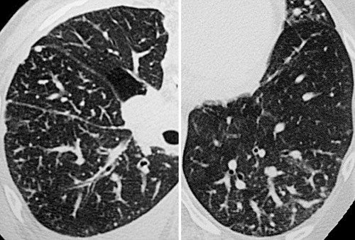
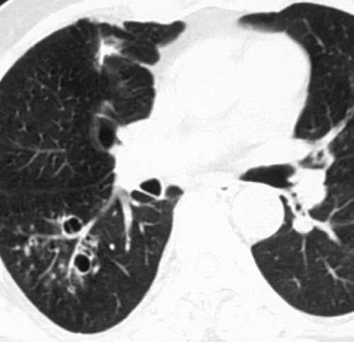
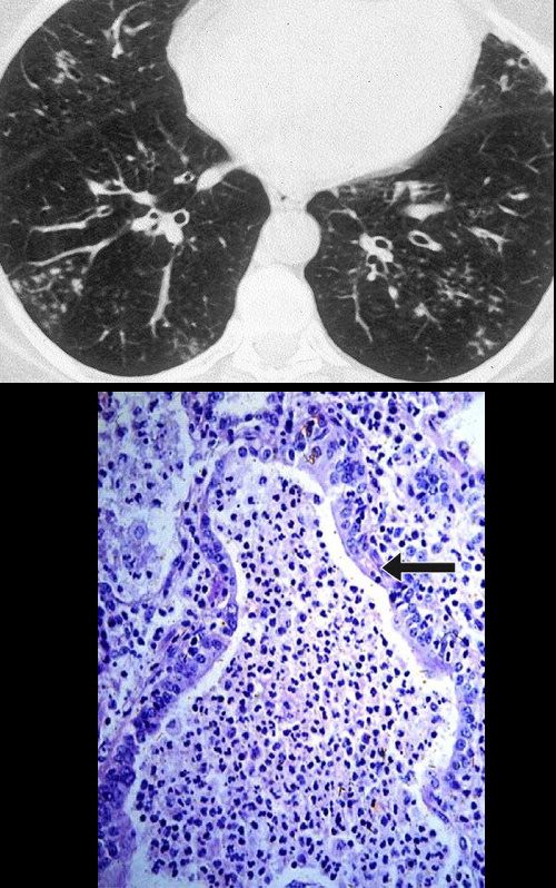
tree in bud opacities in lungs
The purpose of this study was to determine the relative frequency of causes of TIB opacities and identify patterns of disease associated with TIB opacities. 29 Cicatricial scarring of many bronchioles results in the indirect sign of patchy density differences of the lung parenchyma reflecting areas of underventilation and air trapping and subsequent.

Co Rads 2 With Tree In Bud Sign A 27 Year Old Male Attended The Download Scientific Diagram
The tree-in-bud sign reflects the presence of dilated centrilobular bronchioles with lumina that are impacted with mucus fluid or pus.

. 1 5 6 7 8 9 10 11 12. These small clustered branching and nodular opacities represent terminal airway mucous impaction with adjacent peribronchiolar inflammation. Tree-in-bud TIB opacities are a subset of centrilobular nodules.
CT finding of centrilobular nodules with TIB opacities was first described in pulmonary tuberculosis and is considered highly predictive of. Respiratory infections 72 with TB. However to our knowledge the relative frequencies of the causes have not been evaluated.
It is often associated with peribronchiolar inflammation. Usually somewhat nodular in appearance the tree-in-bud pattern is generally most pronounced in the lung periphery and associated with abnormalities of the larger airways. 1 2 3 4 Reported causes include infections aspiration and a variety of inflammatory conditions.
In radiology the tree-in-bud sign is a finding on a CT scan that indicates some degree of airway obstruction. Find the common signs of lung cancer here. The tree-in-bud sign is a nonspecific imaging finding that implies impaction within bronchioles the smallest airway passages in the lung.
Tree-in-bud TIB opacities are a common imaging finding on thoracic CT scan. Multiple causes for tree-in-bud TIB opacities have been reported. TIB opacities typically show branching configurations from secondary pulmonary lobules with sparing of subpleural lungs on CT thorax.
There are several types of GGO. Tree-in-bud TIB opacities are a common imaging finding on thoracic CT scan. These small clustered branching and nodular opacities represent terminal airway mucous impaction with adjacent peribronchiolar inflammation.
A tree-in-bud pattern of centrilobular nodules from metastatic disease occurs by two mechanisms. Alternating areas of normal lung with regions of small airways disease TIB opacities bronchiectasis random small airways pattern was specific 092 for Mycobacterium. Respiratory infections 72 with TB.
As in this case renal cell carcinoma is one of the most common malignancies that may produce this vascular cause of tree-in-bud pattern. Ad Plain English Explanation On The First Sign Of Lung Cancer. Diffuse opacities show up in multiple lobes of one or both lungs.
Nodular opacities with tree-in-bud appearance can be associated with other changes in lung parenchyma-such as thickening of the bronchial walls consolidations andor areas of increased density. Usually somewhat nodular in appearance the tree-in-bud pattern is generally most pronounced in the lung periphery and associated with abnormalities of the larger airways. Multiple causes for tree-in-bud TIB opacities an imaging pattern usually seen on chest CT have been reported.
3 found that the tree-in-bud pattern was seen in. Could you or a loved one have lung cancer. 11 TIB opacities represent a central imag- Background.
1 direct filling of the centrilobular arteries by tumor emboli and 2 fibrocellular intimal hyperplasia due to carcinomatous endarteritis. The tree-in-bud sign is a nonspecific imaging finding that implies impaction within bronchioles the smallest airway passages in the lung. Tree-in-bud refers to a pattern seen on thin-section chest CT in which centrilobular bronchial dilatation and filling by mucus pus or fluid resembles a budding tree.
Multiple causes for tree-in-bud TIB opacities have been reported. Tree-in-bud refers to a pattern seen on thin-section chest CT in which centrilobular bronchial dilatation and filling by mucus pus or fluid resembles a budding tree. However to our knowledge the relative frequencies of the causes have not been evaluated.

Scielo Brazil Tree In Bud Pattern Tree In Bud Pattern

Tree In Bud Caused By Haemophilus Influenzae Radiology Case Radiopaedia Org
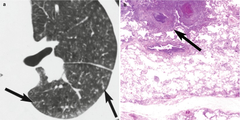
Tree In Bud Sign Radiology Key

Tree In Bud Pattern Pulmonary Tb Eurorad

Tree In Bud Caused By Haemophilus Influenzae Radiology Case Radiopaedia Org
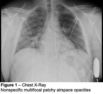
It Is Not Always Tuberculosis Tree In Bud Opacities Leading To A Diagnosis Of Sarcoid Shm Abstracts Society Of Hospital Medicine
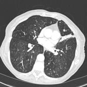
Tree In Bud Sign Lung Radiology Reference Article Radiopaedia Org
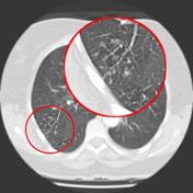
Tree In Bud Sign Lung Radiology Reference Article Radiopaedia Org

A Chest X Ray With A Miliary Pattern And A Tree In Bud Sign Download Scientific Diagram
View Of Tree In Bud The Southwest Respiratory And Critical Care Chronicles
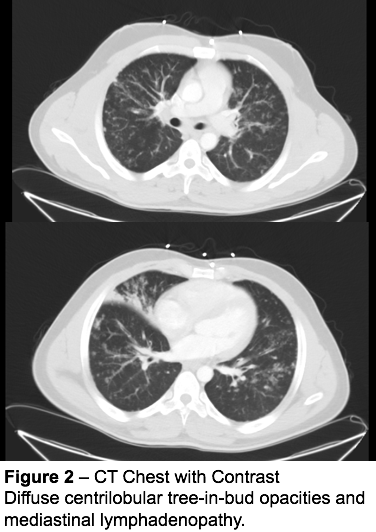
It Is Not Always Tuberculosis Tree In Bud Opacities Leading To A Diagnosis Of Sarcoid Shm Abstracts Society Of Hospital Medicine

Hrct Scan Of The Chest Showing Diffuse Micronodules And Tree In Bud Download Scientific Diagram
